ECG
ECG
What is an ECG?
An Electrocardiogram (ECG or EKG) is a simple, non-invasive test that records the electrical activity of the heart over a period of time. It provides crucial information about the heart's rhythm, structure, and function, making it a vital tool for diagnosing various cardiac conditions.
How Does an ECG Work?
An ECG measures the electrical impulses that trigger each heartbeat. The heart has a natural electrical system that coordinates the contraction of the heart muscle, allowing it to pump blood effectively. When the heart beats, electrical signals travel through the heart muscle, causing it to contract and pump blood. An ECG captures these signals through electrodes placed on the skin.
Components of an ECG Waveform
An ECG waveform consists of several components, each representing a specific phase of the heart's electrical activity:
- P Wave: Represents atrial depolarization (contraction of the atria).
- QRS Complex: Represents ventricular depolarization (contraction of the ventricles). It is typically the most prominent part of the ECG.
- T Wave: Represents ventricular repolarization (recovery of the ventricles).
- U Wave: Sometimes visible, it may represent the repolarization of the Purkinje fibers.
Indications for an ECG
An ECG is commonly performed for various reasons, including:
- Chest Pain: To evaluate the cause of chest pain and rule out heart attacks or other cardiac conditions.
- Irregular Heartbeats: To assess arrhythmias (irregular heart rhythms).
- Pre-Operative Assessments: To evaluate heart health before surgery.
- Monitoring Heart Conditions: For patients with known heart conditions, to monitor changes over time.
- Symptoms of Heart Disease: Such as shortness of breath, fatigue, or palpitations.
Types of ECGs
There are several types of ECGs used for different diagnostic purposes:
- Resting ECG: Conducted while the patient is at rest, typically in a clinic or hospital setting. This is the most common type.
- Stress ECG: Performed while the patient exercises (usually on a treadmill) to assess how the heart responds to physical stress.
- Holter Monitor: A portable ECG device worn by the patient for 24-48 hours to continuously monitor heart activity, useful for detecting intermittent arrhythmias.
- Event Monitor: Similar to a Holter monitor, but worn for a longer period and activated by the patient during symptoms.
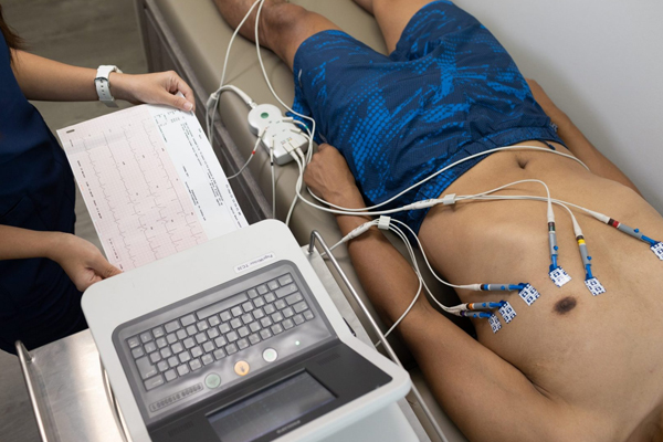
ECG Procedure
The ECG procedure is quick and straightforward:
- Preparation: The patient may be asked to lie down and remove any clothing covering the chest. Skin may be cleaned to ensure good electrode contact.
- Electrode Placement: Small adhesive pads (electrodes) are attached to the chest, arms, and legs. The number and placement of electrodes may vary depending on the type of ECG being performed.
- Recording: The ECG machine is turned on, and it records the electrical signals for a few minutes. The patient is usually asked to remain still and breathe normally during the recording.
- Interpretation: A healthcare professional interprets the ECG results, looking for any abnormal patterns or changes.
Interpreting ECG Results
ECG results are interpreted based on specific patterns and intervals, which can indicate various conditions:
- Normal Sinus Rhythm: Indicates a normal heartbeat.
- Arrhythmias: Irregular heart rhythms, which can be benign or indicate a serious issue.
- Ischemia: Reduced blood flow to the heart, often indicated by changes in the ST segment.
- Myocardial Infarction: Signs of a heart attack, which may show characteristic ST elevation or Q waves.
- Hypertrophy: Enlargement of heart chambers, evident through changes in the waveforms.
Limitations of ECG
While ECGs are powerful diagnostic tools, they have limitations:
- Snapshot of Heart Activity: An ECG reflects the heart's activity only during the time of recording, which may miss intermittent issues.
- False Positives/Negatives: Certain conditions may not be detected, and some non-cardiac factors (like electrolyte imbalances) can cause abnormal results.
- Not Definitive: An abnormal ECG does not always indicate heart disease; further testing may be required for a definitive diagnosis.